Microscopy → interactive 3D
Turn microscope images into explorable 3D specimens.
Create an educational 3D interpretation from one clear reference image.
Invisible structures may be inferred.

Source stays visible
From image to specimen

The source remains attached to the result, so observed evidence and inferred structure are not presented as the same thing.
From a flat photo to a model you can turn around
No modelling software and no waiting — upload an image and get an interactive 3D cell in three steps.
- 1
Upload a microscope photo
Any light- or electron-microscope image of a cell — a textbook figure or your own slide.
- 2
AI identifies the cell
It recognises the cell type and its structures, then builds the matching 3D model in under a minute.
- 3
Explore and share in 3D
Rotate, zoom, annotate, and embed it in slides — or publish it to the community.
From the community
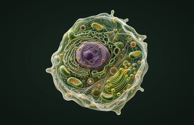
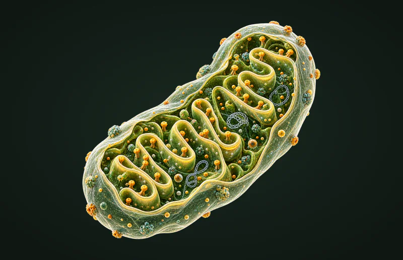
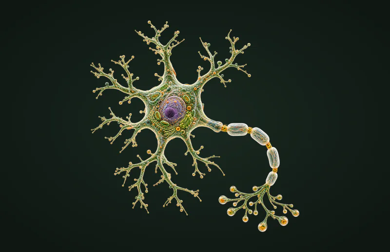
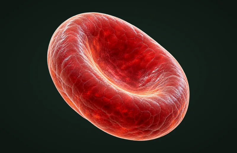
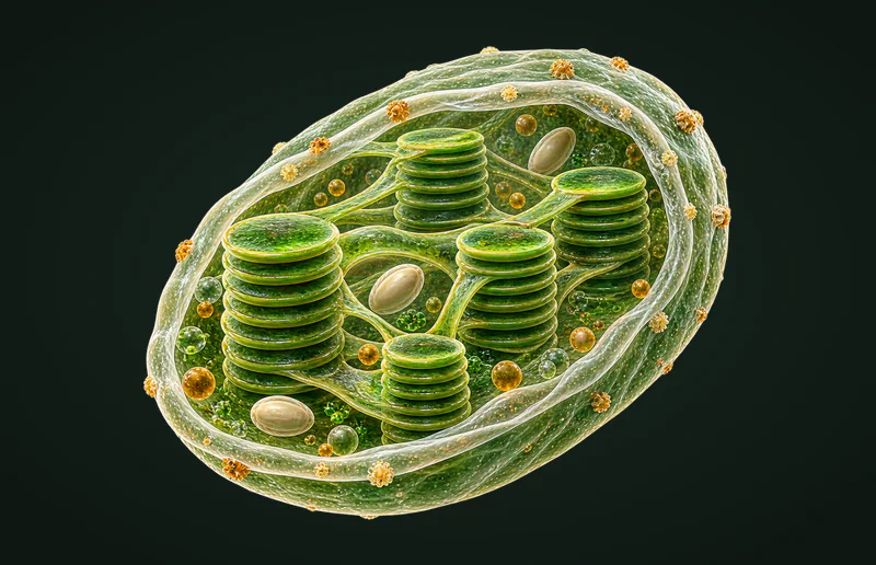
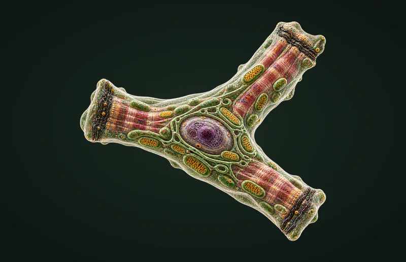
Real 3D cells generated by students like you.
Explore the cell library
30 cells, each with a 3D model and a written guide — structure, function, and exam tips for AP Bio, IB HL, MCAT, and USMLE.
Who it’s for
Students
Turn the flat diagrams in your textbook into something you can rotate and actually understand — then test yourself before the exam.
Teachers
Generate a model from the figure you already teach with, then embed it in your slides or LMS with one snippet.
Curious self-learners
Bring your own microscope photos to life, save them to your library, and share them with the community.
Frequently asked questions
How does Cell Forge turn a 2D photo into a 3D model?
You upload a microscope image; the AI recognises the cell type and its visible structures, then generates an interactive 3D model you can rotate, zoom, and label. It usually takes under a minute.
What kind of images work best?
A clear light- or electron-microscope image where the cell fills most of the frame works best — a textbook figure, a lab slide photo, or a stained section. Blurry or very crowded fields give weaker results.
Which cells and courses are supported?
Thirty cell types — organelles, blood cells, neurons, muscle, epithelial, and more — each mapped to AP Bio, IB HL, A-Level, MCAT, and USMLE topics, with a written guide on every cell page.
Is the 3D model scientifically accurate?
The model is a curriculum-accurate illustration of that cell type — the right structures in realistic proportions — rather than a literal reconstruction of the exact specimen in your photo. Think of it as a teaching figure you can hold and turn.
Can I use it for teaching or put it in my slides?
Yes. Every model has an embed snippet, so you can drop a live, rotatable cell into your slides, website, or LMS. Classroom use is encouraged.
Is it free?
You get one free generation a month on a free account. Pro and Studio plans raise that limit, and credit packs cover one-off bursts. Browsing the cell library and the community gallery is always free.